Molecular MR imaging of molecular and cellular pathways in GBM cell and animal models
Our lab aims at investigating the mechanisms responsible for the resistance of GBM to cancer therapies and to develop strategies to overcome these resistances. For the purpose, we use molecular MR imaging as well as genomics, which make us understand in vitro and in vivo cancer biology regarding metabolism and tumor micro-environment. We are surveying key molecules, cellular pathways and microenvironments, which play an important role for resistance to chemotherapy or anti-angiogenic therapy, and in particular we seek to improve cancer resistance following the therapies by selective inhibition of the molecules, cellular pathways and microenvironments.
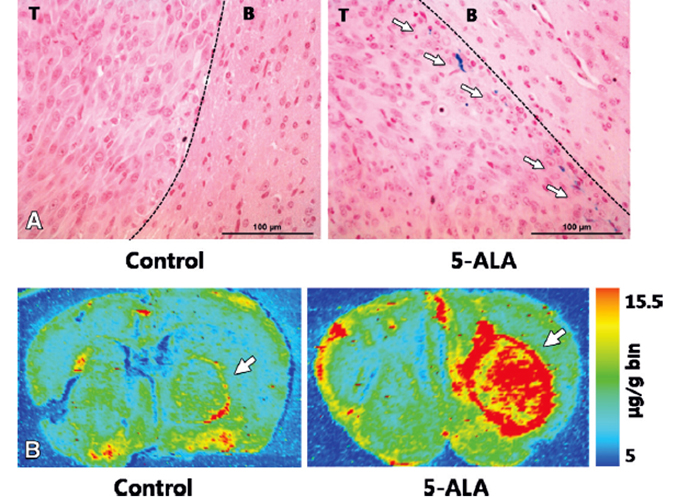
Determination of heme iron in brain tumors at histologic analysis Figure 4: Determination of heme iron in brain tumors at histologic analysis. A, Multifocal iron deposits (arrows) at border (dotted line) between tumor and brain in mice treated with 5-ALA were stained with Prussian blue. No staining was observed in control mice treated with saline. B, Intratumoral iron levels showed substantial difference in mean concentration of iron (arrow) between control animals and 5-ALA–treated animals measured with laser ablation ICP mass spectrometry (38.8 mg/g [IQR, 31–42 mg/g ] vs 51.9 mg/g [48–60.5 mg/g]; P = .014). T = tumor, B = brain.
An NMR metabolomics approach for the diagnosis of leptomeningeal carcinomatosis
Developing new MR diagnostic tools for GBM researches
In both preclinical and clinical researches for GBM, MR imaging is the most important modality for the pre-treatment diagnosis and post-treatment evaluation. Recently, several physiologic and metabolic MR imaging methods are developed and can give several biologic information such as tumor micro-environment and heterogeneity. In our lab, we have a 9.4 T MR scanner for cutting edge preclinical studies, and the preclinical investigations are being transferred to the clinical fields. For the purpose, we are developing new MR diagnostic tools for GBM, which aim at focusing tumor metabolism and several cellular pathways. In addition, we also obtain genomic information from animal model and human GBM for the deep insight of future clinical applications
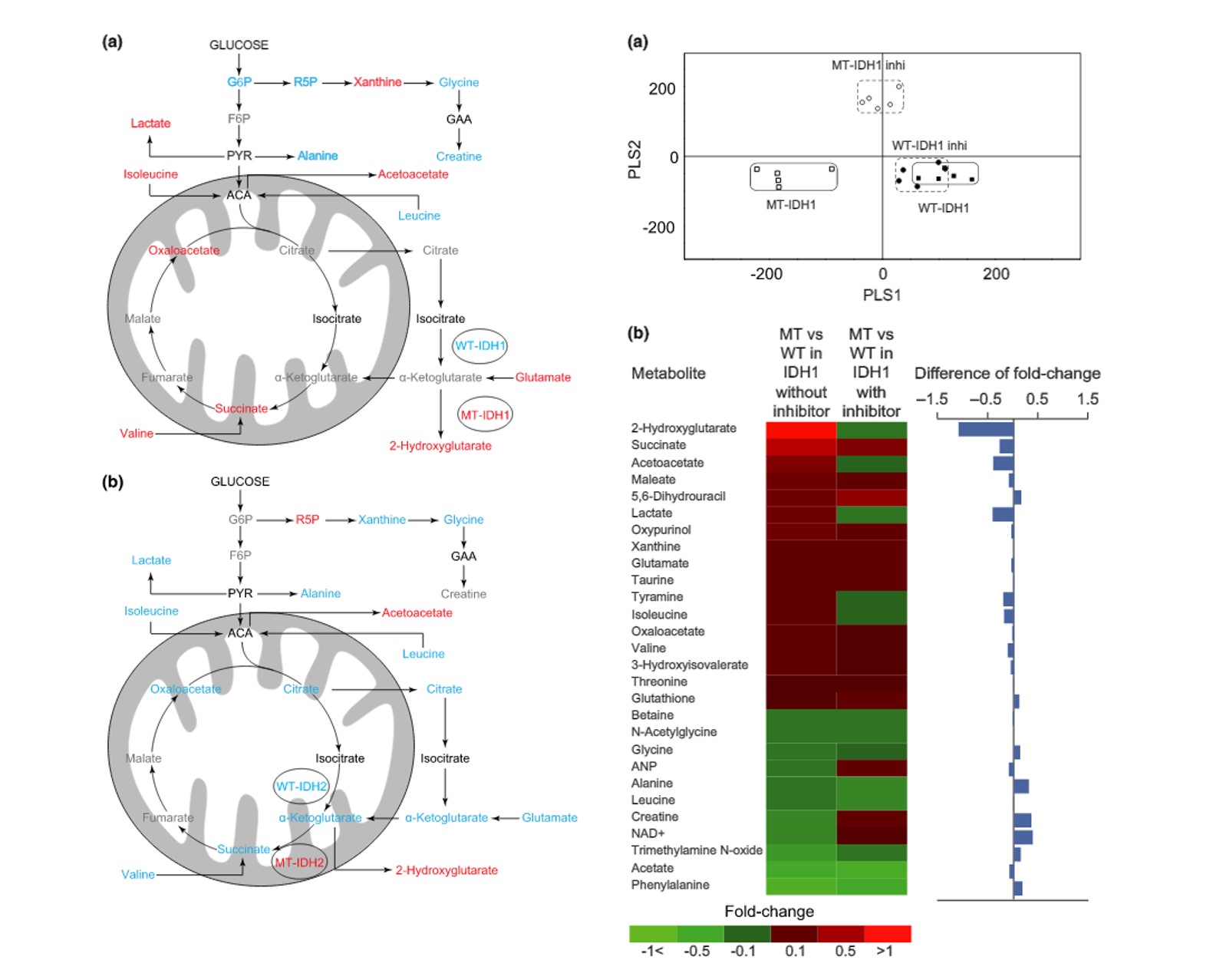
Metabolism research for investigation of IDH mutation subtyping
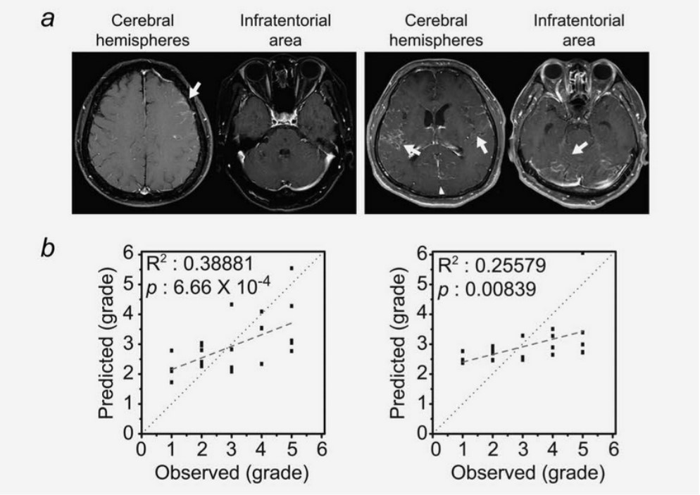
Metabolic profiling and its correlation with MR imaging appearance
Biological application with bio-integrated materials and devices for GBM treatment, and its clinical translation
In terms of treatment for GBM, the standard protocol includes tumor resection followed by concurrent chemoradiotherapy with temozolomide, which was proved to increase patients’ survival. However, many patients suffer from recurrence after completion of the standard therapy, especially around the surgical cavities. So, our lab has been developing biological application with bio-integrated materials and devices for GBM treatment, which mainly aims at suppressing potential malignant cells around the surgical cavities. For the purpose, our lab applies nanotechnology by collaboration with Center for Nanoparticle Research, Institute for Basic Science (IBS).
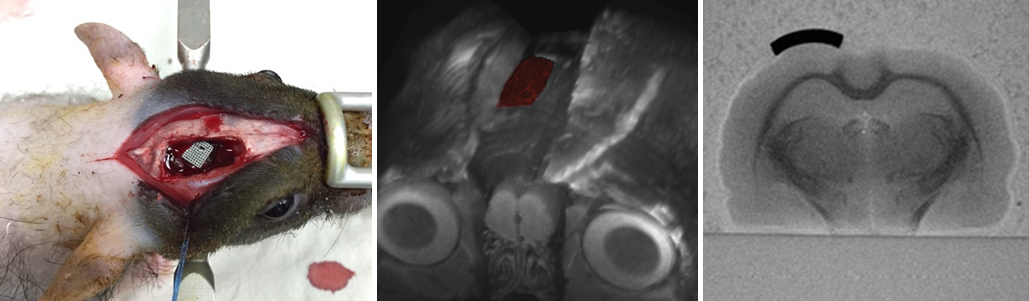
Patch for GBM treatment
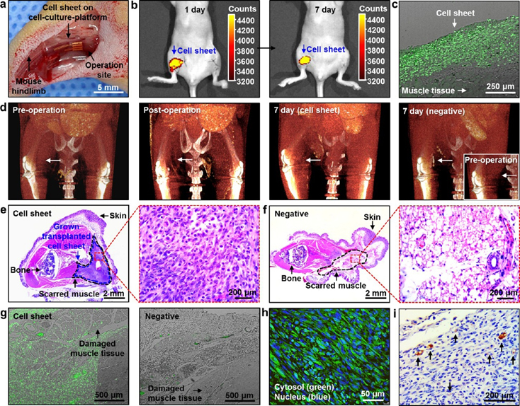
Monitoring for therapeutic effects of cell sheet