Glioblastoma multiforme (GBM) is the most common primary brain tumor in adults; it is also extremely aggressive. GBM is characterized by extensive heterogeneity, angiogenesis, strengthened cell proliferation and cellular invasiveness. Tumor microenvironment of the GBM is very complex due to diverse cell population, and heterogeneous molecular and cellular pathways. For this reason, molecular MR imaging is emerging, which enable to observe different phenomena in the tumor microenvironment and evaluate molecular MR for therapeutic efficacy to anti-cancer drug. In our lab researches, scientists and clinicians work together to translate laboratory findings into new or improved MR diagnostic and therapeutic tool. For the purpose, our lab mainly approaches three aspects of GBMs:
- Molecular MR imaging of molecular and cellular pathways in GBM cell and animal models
- Developing new MR diagnostic tools for GBM researches
- Biological application with bio-integrated materials and devices for GBM treatment, and its clinical translation
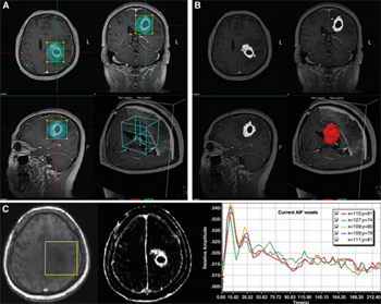
From Tae Jin Yun et al., 2015 Figure 1. Definition of the VOI and acquisition of dynamic contrast-enhanced MR imaging–derived pharmacokinetic parameter maps. A, After the VOI was defined for the enhancing portion, B, the enhancing area was semiautomatically segmented. C, After the identification of the arterial input function (AIF).
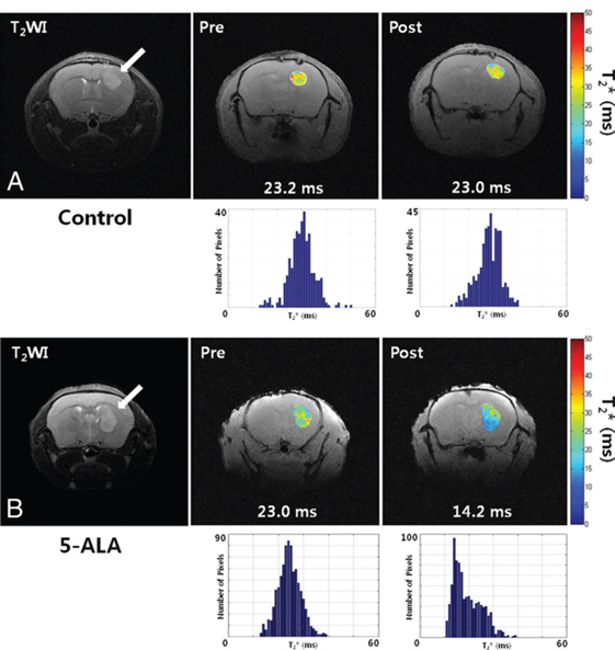
From Hye Rim Cho et al.,2014 Figure 3. T2* maps and graphs show detection of heme synthesized as a result of PpIX generated by 5-ALA treatment in U-87 glioblastomas. Number of pixels (blue bars) was used to calculate median T2* value, and these values were used for detection of hemeinduced changes in T2*. Median T2* value of U-87 glioblastomas treated with 5-ALA was lower than that of control tumors, which received normal saline (P = .011). A, T2* maps showed no significant difference between median T2* values of brain tumors before and after receiving saline (P = .471). B, However, median T2* value of brain tumors significantly decreased after treatment with 5-ALA (P = .031). Arrow indicates U-87 glioblastoma in mouse brain, and numbers are median T2* values of tumors.